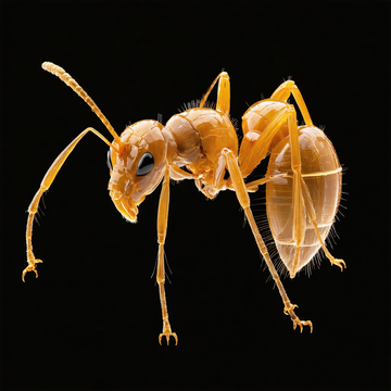
Imagine opening a website and turning an ant around in 3D. You can zoom in, look inside the body, and see muscles, nerves, the gut, and even the stinger. That is the idea behind Antscan, a new open database created by researchers from OIST in Japan, KIT in Germany, and many partners around the world. On March 5, 2026, the team presented the project in Nature Methods. The public portal now offers free interactive scans of about 792 ant species—roughly 800 species—making Antscan the largest online 3D insect database of its kind. (antscan.info)
To build this library, the researchers used synchrotron micro-CT, a very powerful kind of X-ray imaging, at KIT. A robot changed the samples automatically, and the team scanned about 2,000 alcohol-preserved ant specimens in a single week. For each specimen, they captured around 3,000 2D X-ray images and rebuilt them into a detailed 3D model. This method is strong enough to show not only the hard outer shell, but also soft internal tissues that are usually difficult to study. (sciencedaily.com)
AI also plays an important role. The Antscan workflow uses automated processing and artificial intelligence to manage huge amounts of image data. In addition, a follow-up project at the University of Maryland is using AI “pose estimation” to change awkward scan positions into more natural-looking ant shapes. This makes the models easier to study, animate, and use in virtual reality, education, and art. (antscan.info)
Why is this exciting? Antscan is not only beautiful; it is useful. Each entry includes information about taxonomy, ecology, geography, and genome sequencing status. The scans have already helped scientists study how an ant’s body armor connects to colony size and evolution across more than 500 species. Antscan shows how biology, computing, and open data can work together—and how even a tiny ant can open a big new window on life. (antscan.info)